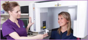
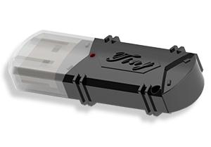
SIERA Dental 3D CT SCAN SOFTWARE Test With sentinel HL dongle
What is ?
CT imaging, or tomography, as it is commonly known among the people, is a method that allows detailed visualization of the anatomical structure of the body. With CT, information about where tumors are located and their size can be obtained. However, it is unclear whether these are malignant or benign lesions.

SIERA Dental 3D Combination of PET and CT
PET/CT is based on combining PET, which produces images of cell metabolism, and CT, which provides anatomical details, in the same device. Thus, both the metabolic function of the cells and the anatomy can be comprehensively visualized by a single device in a single session. Thus, physicians can obtain detailed and precise information about the patient’s anatomical and metabolic status with three-dimensional images. PET/CT enables rapid and accurate diagnosis of especially cancer and heart diseases.
It is possible to determine the structural and functional characteristics of the tumor and to accurately determine. The treatment area, thanks to PET-CT, which is also used to monitor the response to treatment during cancer treatment. Meanwhile, it also allows the healthy tissue around the tumor to be preserved as much as possible. Compared to other planning methods, it significantly reduces the side effects associated with radiotherapy, while providing high doses of radiation to the tumor.
PET/CT is also very valuable in the diagnosis of brain-related diseases, especially dementia (dementia) and epilepsy, and allows early diagnosis of Alzheimer’s disease.
How does the process take place?
Patients who will enter the PET/CT device are first injected with a drug and they have to wait for 45-60 minutes for this drug to be absorbed by the cancer cells that may exist in the body. Then the scan begins and the patient stays in a supine position for 35-45 minutes.


